Thromb Res. 2024 Jun;238:172-183. doi: 10.1016/j.thromres.2024.04.015. Epub 2024 Apr 17.
Fibrin Clot Shields from Cancer Cells: A Barrier to Treatment and Schaffold for Migration
Our research introduces a groundbreaking concept: Cancer cells' procoagulant signature drives the formation of fibrin networks in the tumor microenvironment. Tissue Factor, expressed by cancer cells, plays a key role in shaping the quality and density of these networks.
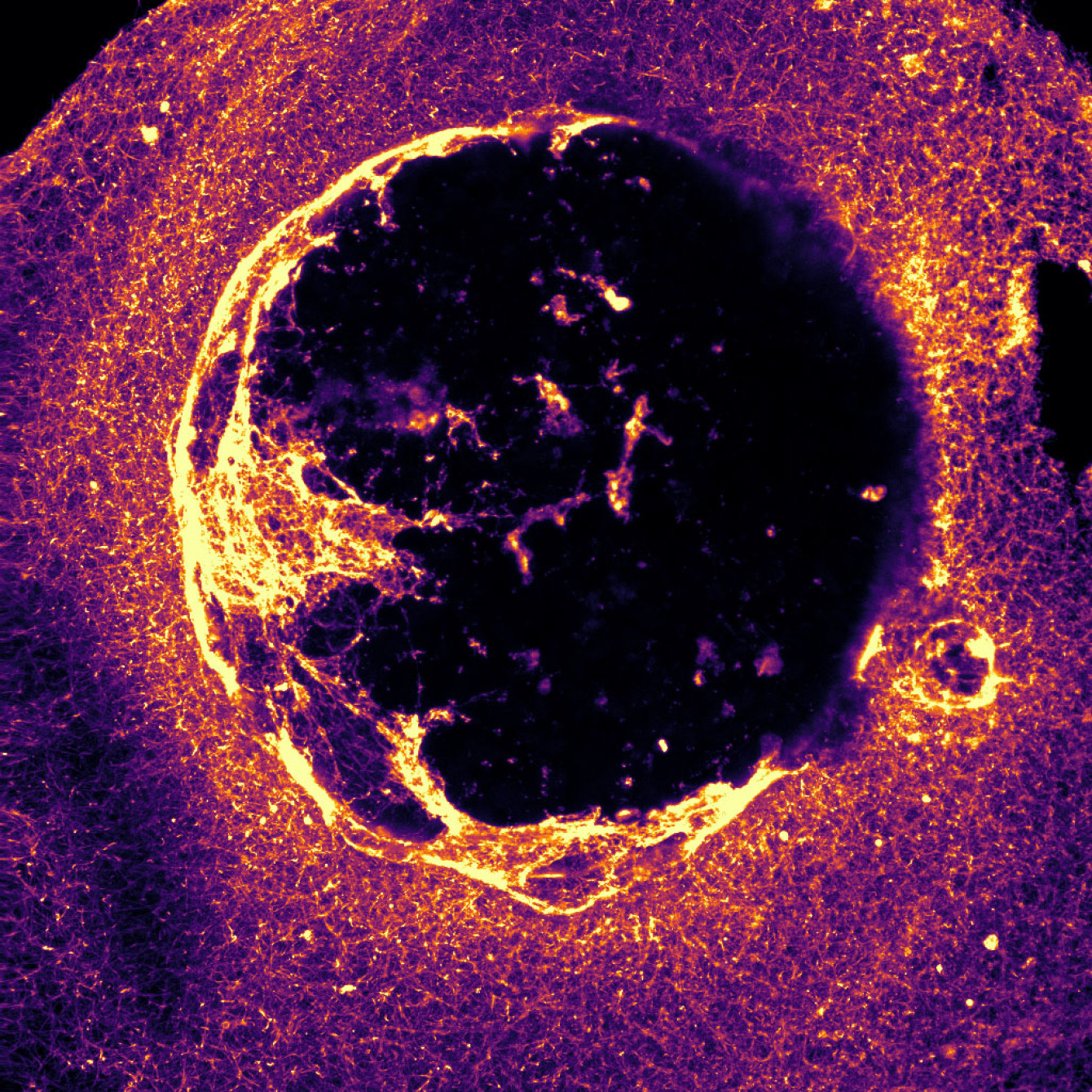
Electron microscopy images vividly show how cancer cells' procoagulant signature leads to the formation of fibrin clot shields. These shields, resembling a "bird's nest," envelop cancer cells, shielding them from anticancer agents and provide a schaffold for migration.
The discovery of thencancer-induced Fibrin Clot Shield sheds light on how the tumor microenvironment contributes to treatment resistance. This highlights the need to identify new therapeutic targets.
Call for Collaboration in the ASTERIX network :
We're forming an international research network to explore this new mechanism of treatment resistance induced by fibrin clot shield formation in the cancer microenvironment.
INTRODUCTION: Cancer cells induce hypercoagulability in the tumoral microenvironment by expressing Tissue Factor (TF). We aimed to study the impact of the procoagulant signature of cancer cells on the quality and structure of fibrin network. We also studied the impact of fibrin clot shield (FCS) on the efficiency of anticancer agents and the migration of cancer cells.
MATERIALS AND METHODS: Pancreatic cancer cells BXPC3 and breast cancer cells MDA-MB231 and MCF7, were cultured in the presence of normal Platelet Poor Plasma (PPP), diluted 10 % in conditioning media. Their potential to induce thrombin generation and their fibrinolytic activity were assessed. The structure of fibrin network was analyzed with Scanning Electron Microscopy (SEM). Cancer cells' mobility with fibrin clot and their interactions with fibrin were observed. Cancer cells were treated with paclitaxel (PTX) or 4-hydroxy-tamoxifen (4OHTam) in the presence or absence of FCS.
RESULTS: Cancer cells, in presence of PPP, induced fibrin network formation. High TF-expressing cancer cells (BXPC3 and MDA-MB23 cells), led to dense fibrin network with fine fibers. Low TF expressing cells MCF7 led to thick fibers. Exogenous TF enhanced the density of fibrin network formed by MCF7 cells. Cancer cells through their inherent profibrinolytic potential migrated within the fiber scaffold. The BXPC3 and MCF7 cells moved in clusters whereas the MDA-MB231 cells moved individually within the fibrin network. FCS decreased the efficiency of PTX and 4OHTam on the viability of cancer cells.
CONCLUSIONS: The procoagulant signature of cancer cells is determinant for the quality and structure of fibrin network in the microenvironment. Original SEM images show the architecture of "bird's nest"-like fibrin network being in touch with the cell membranes and surrounding cancer cells. Fibrin network constructed by triggering thrombin generation by cancer cells, provides a scaffold for cell migration. Fibrin clot shields protect cancer cells against PTX and 4OHTam.

Bâtiment Kourilsky
34 rue Crozatier
75012 PARIS
Sorbonne Université
27 rue Chaligny
75012 PARIS